
Anatomy TUESDAY – Clinical Anatomy Pearl 7 – Fascia
For decades, fascia received little attention in anatomy textbooks.
During dissection, it was often removed simply to expose the structures beneath it. Muscles, tendons, ligaments, nerves, and joints were viewed as the “important” tissues, while fascia was considered little more than connective wrapping.
Today, our understanding has changed dramatically.
Fascia is now recognized as a dynamic, living tissue that contributes to force transmission, proprioception, movement coordination, and even pain perception. While popular media has sometimes exaggerated its role, modern research clearly demonstrates that fascia is far more than anatomical packing material.
For musculoskeletal clinicians, understanding fascia provides another lens through which to view movement dysfunction, injury, and rehabilitation.
What Is Fascia?
Fascia is a continuous network of connective tissue that surrounds, supports, and connects nearly every structure in the body.
It envelops:
- Muscles
- Tendons
- Bones
- Nerves
- Blood vessels
- Organs
- Joint capsules
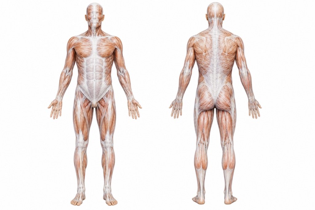
Rather than existing as isolated sheets, fascia forms a continuous three-dimensional matrix extending from head to toe. This continuity explains why movement in one region can influence mechanical behavior elsewhere.
However, fascia should not be viewed as an independent system acting separately from muscles. Instead, muscles and fascia function together as an integrated unit.
Fascia Is Built for Force Transmission
One of the most important discoveries in fascial research is that force does not travel exclusively through tendons. Studies have demonstrated that connective tissue transmits a portion of muscular force both within muscles and between adjacent muscles.
The thoracolumbar fascia provides an excellent example. This dense fascial structure connects the:
- Latissimus dorsi
- Gluteus maximus
- Erector spinae
- Internal oblique
- Transversus abdominis
Located in the lower back, it serves as an attachment site for numerous muscles while contributing to spinal stability. When the gluteus maximus contracts during walking, force is transmitted through the thoracolumbar fascia to the contralateral latissimus dorsi.
This connection forms part of the posterior oblique sling, an important mechanism for rotational stability during gait.
During walking, lifting, and rotational activities, forces generated by the upper and lower extremities are transferred through this fascial network. Rather than acting as passive wrapping, the fascia contributes to efficient load distribution throughout the body.
Fascia Is Richly Innervated
One reason fascia has become increasingly important in pain science is its abundant sensory innervation.
Research has identified numerous mechanoreceptors and free nerve endings within fascial tissue.
These receptors respond to:
- Mechanical deformation
- Pressure
- Stretch
- Movement
Inflammation or injury involving fascia may therefore contribute to pain independent of muscles or joints. This helps explain why patients sometimes report diffuse, poorly localized discomfort that cannot be fully explained by imaging findings.
Fascia and Tendons Are Partners
Fascia and tendons share similar collagen-based architecture but serve different functions.
Tendons primarily transmit force from muscle to bone. Fascia distributes force among multiple structures while allowing tissues to glide relative to one another. Healthy fascial mobility reduces friction between muscles during movement.
When tissues lose mobility due to injury, surgery, or prolonged immobilization, normal gliding may be impaired. The result is increased mechanical resistance during movement.
Fascia Responds to Mechanical Loading
Like tendon and bone, fascia is a living tissue capable of adaptation.
Appropriate loading stimulates collagen remodeling and helps maintain tissue health. Conversely, prolonged inactivity may reduce tissue stiffness, alter hydration, and diminish mechanical efficiency.
Regular movement—including strength training, walking, and dynamic mobility exercises—likely provides sufficient stimulus for healthy fascial adaptation in most individuals.
While stretching and manual therapy may temporarily alter tissue behavior or improve comfort, long-term changes in fascial properties appear to depend primarily on progressive loading and habitual movement.
Why does this matter?
Fascia is not merely connective wrapping. It is a dynamic tissue that contributes to force transmission, proprioception, movement coordination, and pain perception. Appreciating its role helps clinicians better understand regional interdependence and reinforces the importance of treating movement systems rather than isolated anatomical structures.
Don’t just ask, “How do I release the fascia?” Ask, “How can I improve the way this movement system loads, transfers force, and adapts?” The answer usually lies not in chasing fascial restrictions but in restoring efficient, progressive movement throughout the entire kinetic chain.
Join me next week as we explore peripheral nerve anatomy.
Because nobody has time to be in pain.
Until next time…

Kind Regards,
MoveWell Academy
[email protected]