
Kinesiology THURSDAY – Biomechanics of the Head Turn

Do this test right now. Turn your head to the left as far as you can. Then turn your head to the right as far as you can.
Do those feel the same or is one direction more limited? Do you feel pain or discomfort in end range? Do you hear crackling noises?
Turning your head to check your blind spot while driving seems effortless. Looking over your shoulder during a conversation or following a tennis ball across the court happens almost automatically. Yet cervical rotation is one of the most sophisticated movements in the human body.
Neck rotation requires the precise coordination of seven cervical vertebrae, dozens of muscles, multiple ligaments, and the nervous system. When even one part of this system becomes restricted, overloaded, or poorly coordinated, neck pain and limited motion often follow.
Osteokinematics of the head turn
The cervical spine consists of seven vertebrae divided into two functional regions.
The upper cervical spine includes the atlas (C1) and axis (C2), which specialize in rotation. The lower cervical spine (C3–C7) contributes rotation while also allowing flexion, extension, and lateral flexion.
Together these segments produce approximately 70–90 degrees of rotation to each side, although normal values vary with age and measurement technique.
One of the most remarkable facts about cervical motion is that about half of all cervical rotation occurs at a single joint—the atlantoaxial (C1–C2) joint.
The atlantoaxial joint functions much like a pivot.
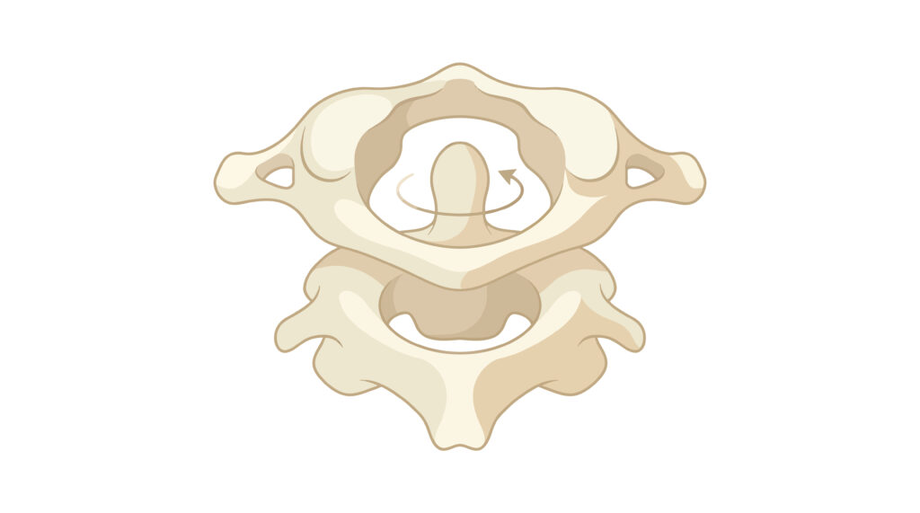
The dens (odontoid process) of C2 acts as the central axis around which the atlas rotates. Strong ligaments—including the transverse ligament and alar ligaments—maintain stability while allowing remarkable mobility.
Approximately 35–45 degrees of rotation occur at C1–C2 in each direction, representing nearly 50% of total cervical rotation. This unique anatomy allows large amounts of rotation while protecting the spinal cord and vertebral arteries.
The remaining cervical rotation occurs between C2 and C7.
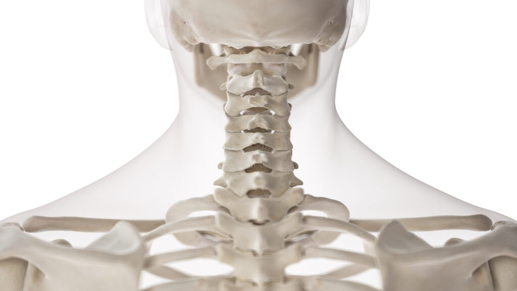
Unlike C1–C2, each lower cervical motion segment contributes only a small amount of movement—typically 5 to 8 degrees. Individually these movements appear insignificant, but collectively they account for the remaining half of cervical rotation.
These segments also allow flexion, extension, and side bending, making them far more versatile than the upper cervical spine.
Coupled movement
One of the most misunderstood aspects of cervical biomechanics is that pure rotation rarely occurs in the lower cervical spine.
Because the cervical facet joints are oriented approximately 45 degrees between the frontal and transverse planes, rotation naturally couples with side bending.
During right rotation:
- The lower cervical vertebrae also side bend to the right.
- The facet joints on the right approximate.
- The facet joints on the left separate.
- Small translational movements occur between adjacent vertebrae.
This coupled motion allows smooth, efficient movement while distributing forces across multiple joints. This also means if you have limited side bending (ear-to-shoulder movement), you will also have limited rotation.
The upper cervical spine behaves differently. At C1–C2, rotation occurs with minimal side bending because of the joint’s unique pivot design.
The muscles behind rotation
Neck rotation depends on both local stabilizers and larger movement-producing muscles.
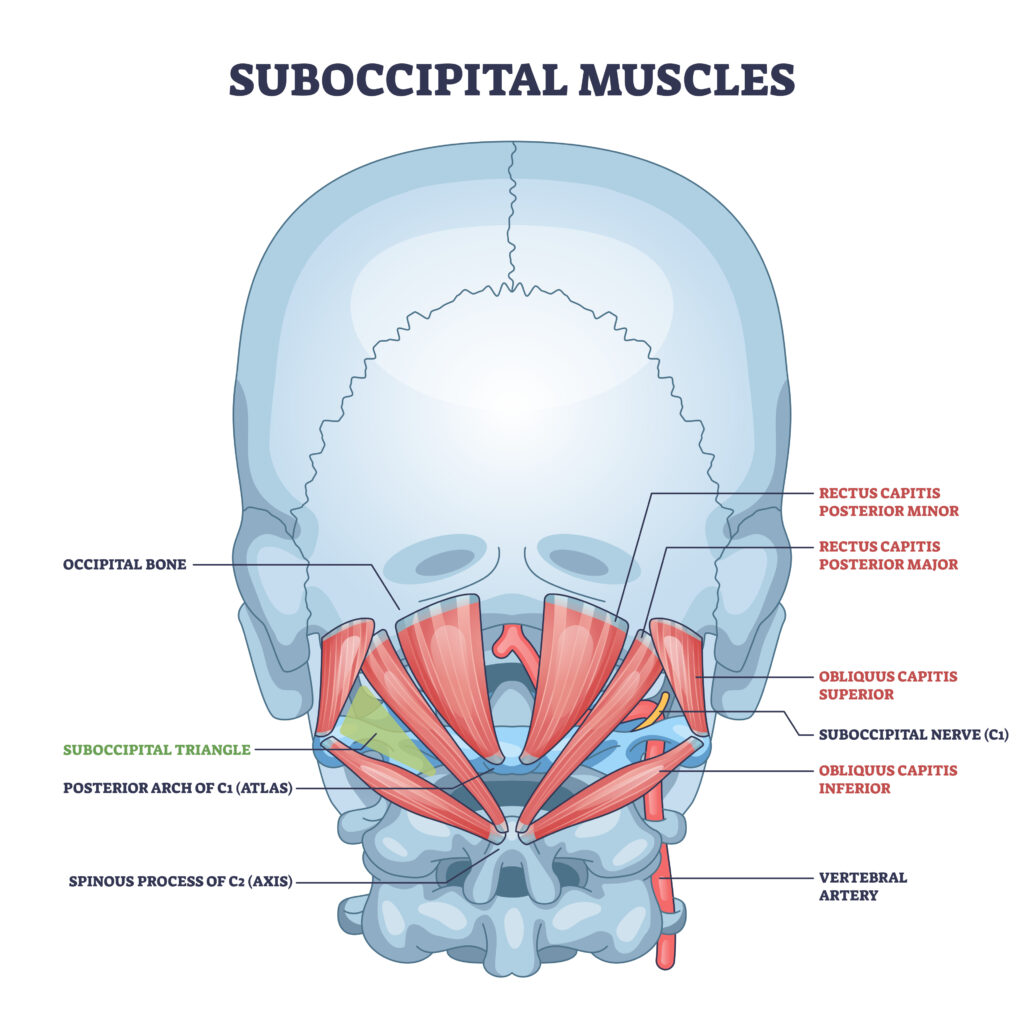
Primary rotators include:
- Sternocleidomastoid
- Splenius capitis
- Splenius cervicis
- Semispinalis capitis
- Multifidus
- Levator scapulae
- Upper trapezius
- Obliquus capitis inferior
- Rectus capitis posterior (major and minor)
Interestingly, the sternocleidomastoid rotates the head to the opposite side, while the splenius muscles rotate to the same side. These muscles must work together with the deep cervical stabilizers to produce smooth, controlled movement.
Trigger points in any of the above muscles will alter the kinematics of the head turn.
The role of the thoracic spine
Many clinicians focus exclusively on the neck when rotation becomes limited. However, cervical rotation depends heavily on thoracic mobility.
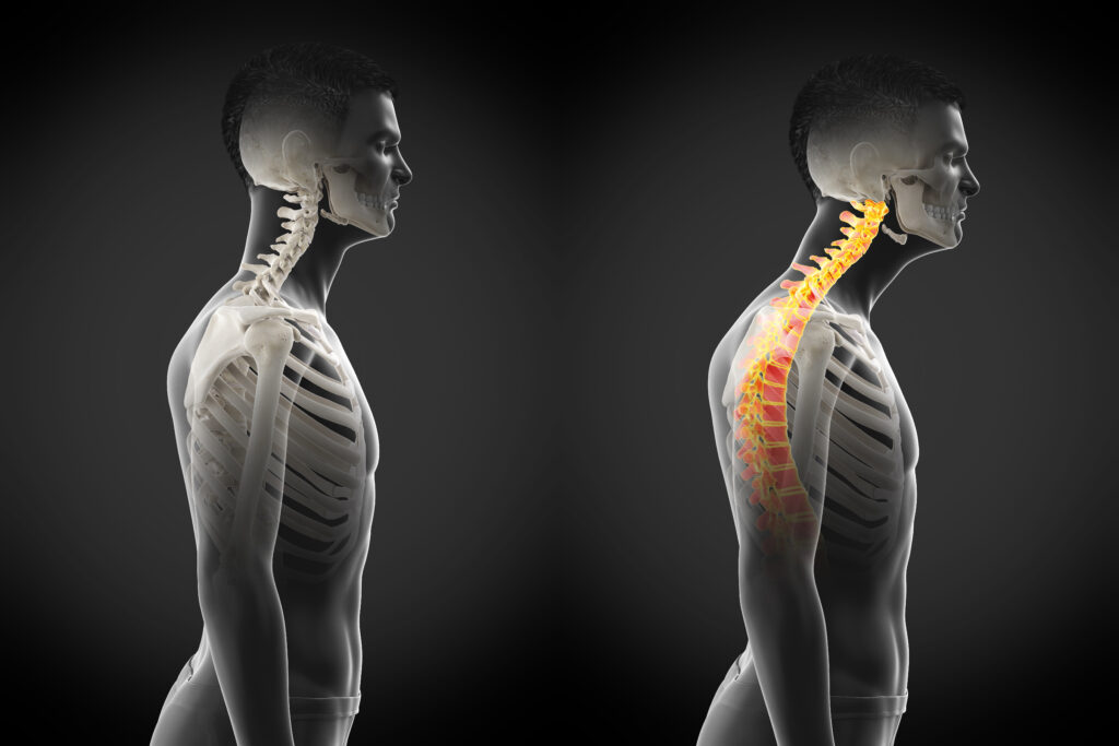
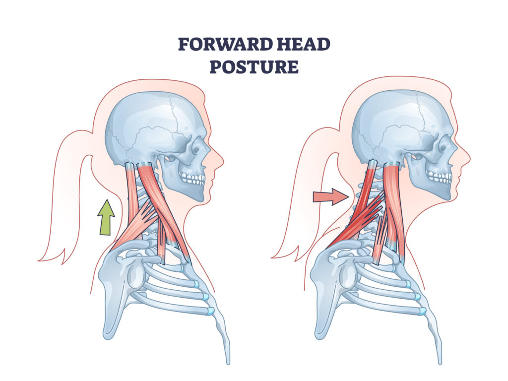
With prolonged forward head posture, the upper cervical spine adapts by extending. This increases compression of the atlanto-occipital joints and tension in the anterior structures, possibly limiting rotation. It also requires increased muscle forces to hold your head on your shoulders, creating muscle fatigue and/or trigger points.
If the upper thoracic spine becomes stiff, the cervical spine often compensates by moving excessively. This increased demand may overload cervical joints and muscles, contributing to pain and reduced motion.
Improving thoracic extension and rotation frequently restores cervical movement without aggressive treatment directed at the neck itself.
Why does this matter?
Turning your head is far more complex than it appears. It requires the coordinated interaction of the upper cervical pivot joint, the lower cervical facet joints, the thoracic spine, and a highly organized network of muscles working together to balance mobility with stability.
When one part of this system loses mobility or control, the rest of the cervical spine compensates. Understanding these biomechanical relationships allows clinicians to identify the true source of movement dysfunction and develop treatment strategies that restore efficient, pain-free motion rather than simply addressing symptoms.
So, turn your head left and right. Make sure you still can.
Because nobody has time to be in pain.
Until next time…
Kind Regards,
MoveWell Academy
[email protected]