Anatomy TUESDAY – Radial Nerve
A patient presents with pain along the outside of the elbow. Gripping hurts. Resisted middle-finger extension reproduces symptoms. The obvious diagnosis may seem to be lateral epicondylitis.
But what if the tendon isn’t the primary problem?
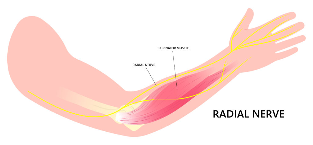
The radial nerve is one of the great mimickers in orthopedic practice. Because it travels from the brachial plexus through the posterior arm, around the humerus, across the lateral elbow, and into the forearm and hand, irritation along its course can resemble shoulder pathology, lateral elbow tendinopathy, or even cervical radiculopathy.
For physical therapists, understanding the path of the radial nerve can dramatically improve differential diagnosis and treatment.
The path of the radial nerve
The radial nerve originates from the posterior cord of the brachial plexus, receiving contributions primarily from C5–T1.
From the axilla, it travels posteriorly with the profunda brachii artery and wraps around the humerus in the radial (spiral) groove.
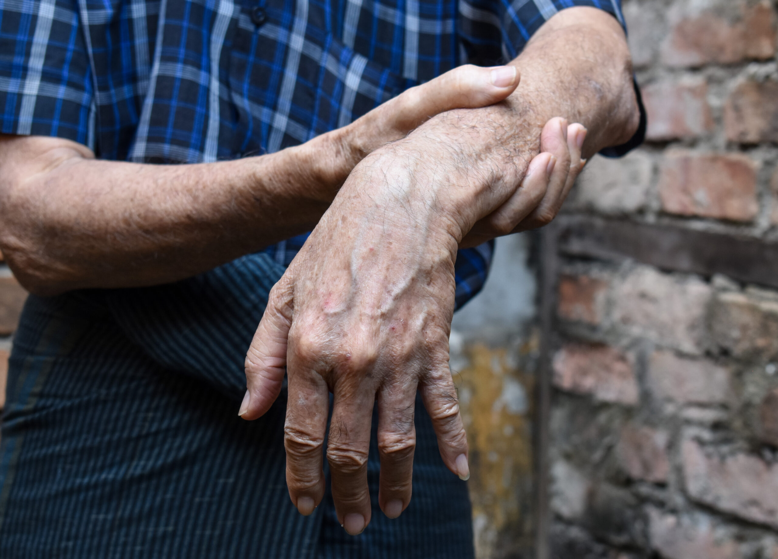
This relationship has an important clinical consequence: a humeral shaft fracture can injure the radial nerve and produce weakness of wrist and finger extension—classically presenting as wrist drop.
As the nerve approaches the elbow, it moves anterior to the lateral epicondyle and divides into two major branches:
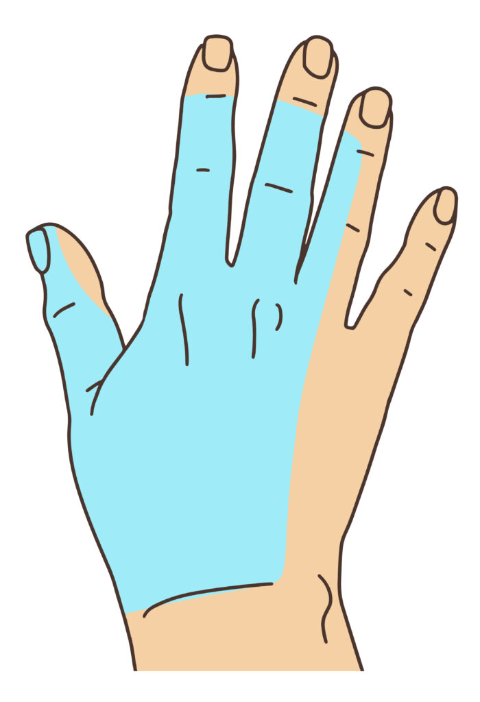
- The superficial radial nerve, primarily sensory to the thumb side of the wrist, back of the hand and thumb or index finger
- The deep radial nerve, primarily motor, which continues through the supinator and becomes the posterior interosseous nerve (PIN)
That division is particularly important when evaluating lateral elbow and proximal forearm pain.
Radial tunnel syndrome is primarily a pain syndrome. Patients generally do not demonstrate clear motor paralysis.
Posterior interosseous nerve syndrome, on the other hand, is primarily a motor neuropathy. Patients may develop weakness of finger and thumb extension, while cutaneous sensory loss is absent because the PIN does not provide cutaneous sensation.
Radial Tunnel Syndrome v. Tennis Elbow
With lateral epicondylitis, maximal tenderness is generally close to the common extensor tendon at the lateral epicondyle.
With radial tunnel syndrome, tenderness is typically farther distal, over the proximal radial forearm along the radial tunnel.
Resisted middle-finger extension and resisted supination may provoke radial-tunnel-region pain, but neither test should be considered diagnostic by itself.
This is an important clinical pearl:
A provocative test tells you that you’ve reproduced symptoms. It doesn’t automatically tell you which tissue caused them.
Palpation, symptom distribution, strength, neurological findings, cervical screening, neurodynamic testing, and the patient’s history should all contribute to the diagnosis.
What does the radial nerve do?
The radial nerve is closely associated with the upper extremity’s extensor system.
Its branches contribute to:
- Elbow extension
- Wrist extension
- Finger extension
- Thumb extension and abduction
- Forearm supination
The superficial radial nerve also supplies sensation to part of the dorsolateral hand.
This means radial nerve dysfunction can present very differently depending on where along the nerve the problem occurs.
A proximal lesion may affect the triceps, wrist extensors and finger extensors.
A lesion around the radial groove may spare much of the triceps but weaken wrist and finger extension.
A PIN lesion primarily affects finger and thumb extension.
A superficial radial nerve problem produces sensory symptoms without motor weakness.
Localization matters more than simply identifying “radial nerve involvement.”
Glide and tension
Like the median nerve, the radial nerve is not a stationary electrical cable.
It must move relative to surrounding muscles, fascia, bones and joints as the shoulder, elbow, forearm and wrist change position.
Clinicians can progressively increase mechanical loading of the radial nerve through combinations of:
Shoulder depression → internal rotation → elbow extension → forearm pronation → wrist flexion/ulnar deviation, with cervical movement used for structural differentiation.
Here’s a link to some great radial nerve glides and tensioners.
Why does this matter?
The radial nerve should be on every orthopedic clinician’s differential when evaluating persistent lateral elbow or proximal forearm pain. Its close relationship with the extensor musculature means neural and tendon disorders can produce remarkably similar symptoms. Know the anatomy well enough to follow the nerve.
Join me next week as we discuss the ulnar nerve.
Because nobody has time to be in pain.
Until next time…

Kind Regards,
MoveWell Academy
[email protected]