Anatomy TUESDAY – The First Rib: A Small Rib with a Big Job
When we think about ribs, we usually think about breathing and protecting the lungs.
But the first rib is different.
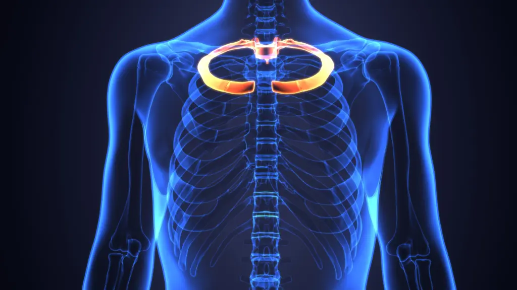
Hidden underneath the clavicle at the base of the neck, the first rib sits at a busy anatomical crossroads. Muscles attach to it. Blood vessels travel across it. The brachial plexus passes just above it. And its position changes as we breathe.
In fact, some of the most important structures traveling from the neck into the arm must negotiate their way around the first rib.
The First Rib Is Different From the Others
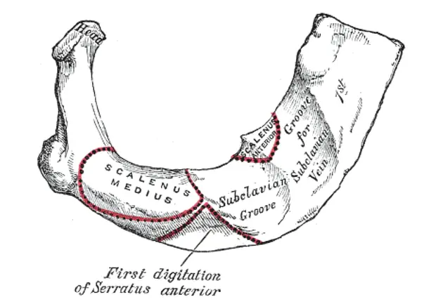
Superior view of the first rib (Gray’s Anatomy)
The first rib is the shortest, broadest, flattest and one of the most curved ribs.
Posteriorly, its head articulates with the body of the T1 vertebra, while anteriorly it connects through its costal cartilage to the manubrium.
Unlike most ribs, which have relatively obvious superior and inferior borders, the first rib is oriented more horizontally and has distinct features on its superior surface.
One of the most important is the scalene tubercle.
And this little bump helps organize much of the anatomy surrounding the first rib.
The Scalene Tubercle Is an Anatomical Traffic Divider
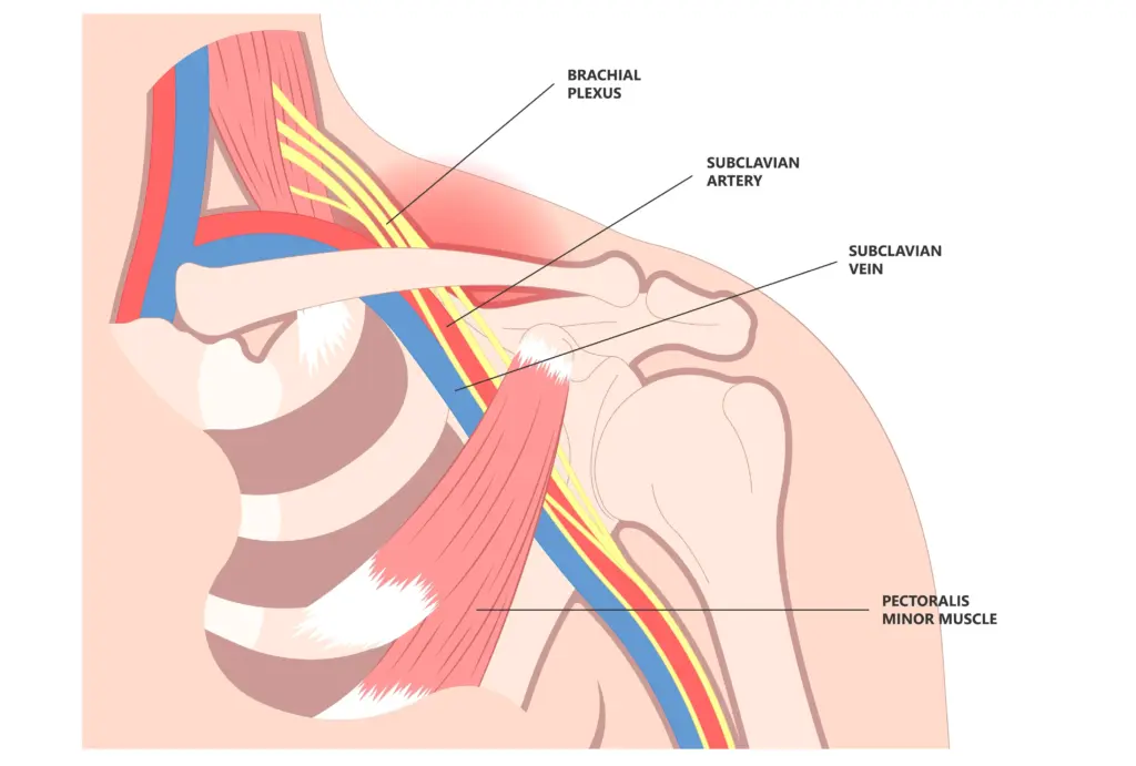
The anterior scalene muscle descends from the cervical spine and attaches to the scalene tubercle of the first rib.
But look at what passes on either side of it.
The subclavian vein crosses over the first rib anterior to the anterior scalene. The subclavian artery and brachial plexus pass in between the anterior and middle scalene muscles.
The space between the anterior scalene, middle scalene and first rib is called the interscalene triangle. The first rib forms its floor beneath the nerves and blood vessels supplying the upper extremity.
This creates a remarkably crowded region.
The First Rib Moves When You Breathe
The first rib isn’t stationary.
The anterior and middle scalene muscles can elevate the first rib and act as accessory muscles of inspiration.
During quiet breathing, the diaphragm does most of the work. But as respiratory demand increases, the scalenes can assist by elevating the upper ribs and helping expand the upper thorax.
The costoclavicular space
The neurovascular structures aren’t finished navigating tight spaces once they leave the scalene triangle. They next travel through the costoclavicular space, the space between the clavicle and first rib.
That means the available space for these structures isn’t determined by one bone alone. It depends on the relationship among the first rib, clavicle, surrounding muscles and soft tissues. And these relationships are dynamic.
Moving the arm, shoulder girdle, neck or thorax can change the dimensions of these spaces.
What Does This Have to Do With Thoracic Outlet Syndrome?
The anatomy of the first rib becomes especially important when discussing thoracic outlet syndrome (TOS).
Thoracic outlet syndrome describes a group of conditions involving compression of neural or vascular structures as they travel from the neck toward the upper extremity.
Potential compression can occur in several anatomical regions, including the:
- Interscalene triangle
- Costoclavicular space
- Pectoralis minor/subcoracoid region
The first rib contributes directly to the boundaries of the first two.
Anatomical variations—including abnormal first ribs, cervical ribs, fibrous bands or variations in scalene anatomy—can potentially reduce the available space for the brachial plexus or subclavian vessels.
But there is an important clinical point here:
Having tenderness, muscle tightness or an apparently “elevated first rib” does not automatically mean someone has thoracic outlet syndrome.
TOS can be diagnostically challenging, particularly neurogenic TOS. Symptoms, examination findings, anatomical relationships and other potential causes of upper-extremity symptoms all need to be considered.
The anatomy helps us understand what could happen. It doesn’t make the diagnosis by itself.
An elevated first rib
Clinicians frequently use the term elevated first rib.
Anatomically, elevation of the first rib is certainly possible.
But clinically, cervical movement, breathing mechanics, scalene activity, clavicular and scapular movement, neural sensitivity, vascular findings and the patient’s symptoms should be considered rather than just treating the first rib as an isolated structure that simply needs to be “put back down.
Some things that may “elevate” a first rib:
- Trigger points in the anterior and middle scalene
- An elevated scapula (upper trapezius and levator scapula)
- An elevated pelvis on the same side (quadratus lumborum)
Why does this matter?
The first rib provides a perfect example of why regional anatomy matters. The first rib is more than part of the rib cage. It forms the floor of the interscalene triangle and the inferior boundary of the costoclavicular space, placing it directly beneath the nerves and blood vessels traveling from the neck into the arm.
A patient experiencing symptoms into the arm may have structures involved that are nowhere near the location where the symptoms are actually felt.
So when evaluating upper-extremity symptoms, the first rib reminds us to look beyond the shoulder, elbow or hand. Sometimes the important anatomy is hiding underneath the clavicle.
Consider the first rib.
Because nobody has time to be in pain.
Until next time…

Kind Regards,
MoveWell Academy
[email protected]