Anatomy TUESDAY – Clinical Anatomy Pearls Every MSK Clinician Should Know
In an era of advanced imaging, evidence-based rehabilitation, and evolving pain science, it can be tempting to view anatomy as a foundational science best left in the classroom. However, for musculoskeletal (MSK) clinicians, anatomy remains one of the most valuable tools for clinical reasoning, differential diagnosis, and patient education.
The difference between a good clinician and a great clinician often lies not in memorizing every anatomical structure, but in understanding how anatomy influences function, dysfunction, and treatment outcomes. Over the next few weeks, we’ll take a dive into several foundational pearls of anatomy wisdom. Join me for the next seven weeks as we discuss the following:
Pain Referral Patterns (Listen with your ears, then take your eyeballs and look elsewhere)
Patients often localize pain inaccurately. Understanding referral patterns is critical for accurate diagnosis. For example, pain felt in the lateral shoulder may originate from cervical nerve root irritation rather than the rotator cuff. Similarly, hip pathology frequently presents as groin pain, but can also refer to the anterior thigh or knee.
The Rotator Cuff (More Than Four Muscles)
Most clinicians can recite the four rotator cuff muscles and their actions, but their functional role extends far beyond shoulder movement.The rotator cuff acts as a dynamic stabilizing system, compressing the humeral head into the glenoid while larger muscles generate movement. Dysfunction often results not from weakness alone, but from altered force coupling between the cuff and surrounding musculature. And weakness is not usually weakness, but overload.
Not All Tendons Are Created Equal
Tendon anatomy varies significantly throughout the body. Differences in vascularity, collagen organization, and mechanical loading influence both injury risk and healing potential.
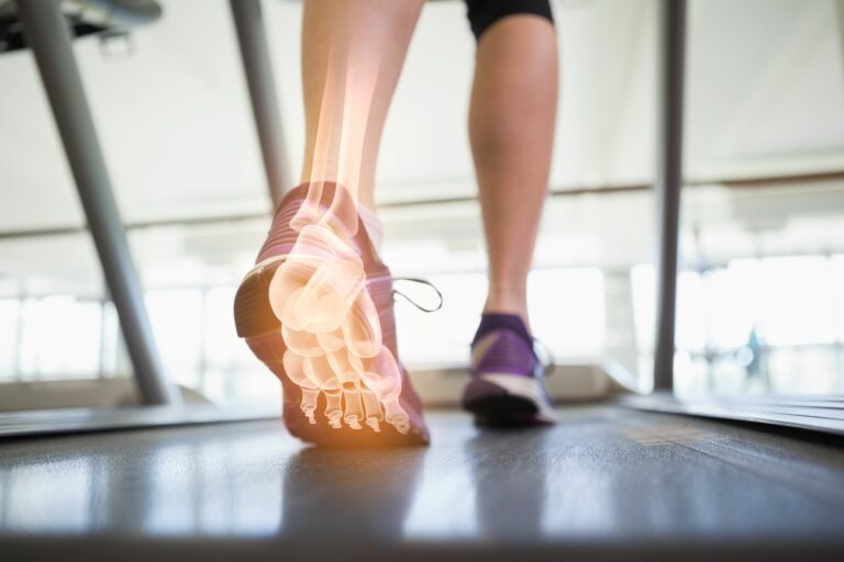
The Achilles tendon, for example, contains a relatively hypovascular region approximately 2–6 cm proximal to its insertion, making it particularly susceptible to tendinopathy. Likewise, the supraspinatus tendon experiences substantial compressive and tensile loads that affect tissue adaptation.
No Man is An Island (And Neither is The Lumbar Spine)
Low back pain remains one of the most common complaints encountered in MSK practice. Yet clinicians who focus exclusively on lumbar anatomy may miss contributing factors elsewhere. Thoracic spine, hip and ankle mobility have significant impact on lumbar function.
Follow Me (Peripheral Nerves Pathways)
Knowledge of peripheral nerve anatomy remains one of the most underutilized clinical skills. Median nerve symptoms may mimic cervical radiculopathy. Peroneal nerve irritation can resemble lumbar pathology. Ulnar nerve entrapment may be mistaken for medial elbow conditions.
Understanding nerve pathways, common entrapment sites, and sensory distributions allows clinicians to perform more targeted examinations and avoid misdiagnosis.
The Scapula Is the BUTT of the Upper Extremity Function
The scapula serves as the base for shoulder movement and force production. Even subtle alterations in scapular position or movement can influence glenohumeral mechanics.
Weakness, fatigue, pain, or altered motor control of the serratus anterior and lower trapezius may contribute to inefficient movement patterns and symptom development. Clinicians should understand how to assess scapular control during functional tasks rather than relying solely on static posture observations.
Teach it to Your Grandma (Anatomy Improves Patient Education)
Finally, this is my favorite. One of the most powerful applications of anatomy is communication. In all my years of teaching, I have encountered many students who were able to recite anatomy facts from a textbook. But a true understanding of anatomy means you are able to turn complex ideas into simple language.
Patients often arrive with misconceptions about their condition. A simple explanation of how muscles, tendons, joints, or nerves function can improve understanding, reduce fear, and increase adherence to treatment plans.
We can use our knowledge of anatomy to empower patients and remove fear around structural findings and test results.
Why does this matter?
Modern MSK care extends far beyond anatomy alone. Pain science, psychosocial factors, and movement behavior all contribute to patient outcomes. However, anatomy remains the framework upon which effective assessment and treatment are built.
The most successful clinicians do not simply memorize structures—they understand how anatomy influences movement, guides differential diagnosis, and shapes clinical decision-making. As healthcare continues to evolve, anatomy remains as relevant as ever. Not because it provides all the answers, but because it helps clinicians ask better questions.
Anatomical knowledge is most valuable when it moves beyond memorization and becomes a tool for clinical reasoning. Every patient encounter presents an opportunity to connect structure, function, and symptom presentation in a meaningful way—and that is where anatomy truly comes alive.
At the end of this series, I hope you are inspired to take the basics of what you learned so many years ago and apply it to the real world: to guide your evaluation and to help your patients understand. You may already know what I am about to teach, but repetition breeds impression. Let’s get back to the basics.
Because nobody has time to be in pain.
Until next time…

Kind Regards,
MoveWell Academy
[email protected]