

Anatomy TUESDAY – Clinical Anatomy Pearl 8 – Peripheral Nerves
Patients rarely walk into the clinic saying, “I think my radial nerve is irritated.”
Instead, they say:
“My shoulder aches.“
“My hand goes numb at night.”
“My elbow hurts every time I lift something.“
“I have burning pain down my leg.”
For musculoskeletal clinicians, these complaints present an important challenge.
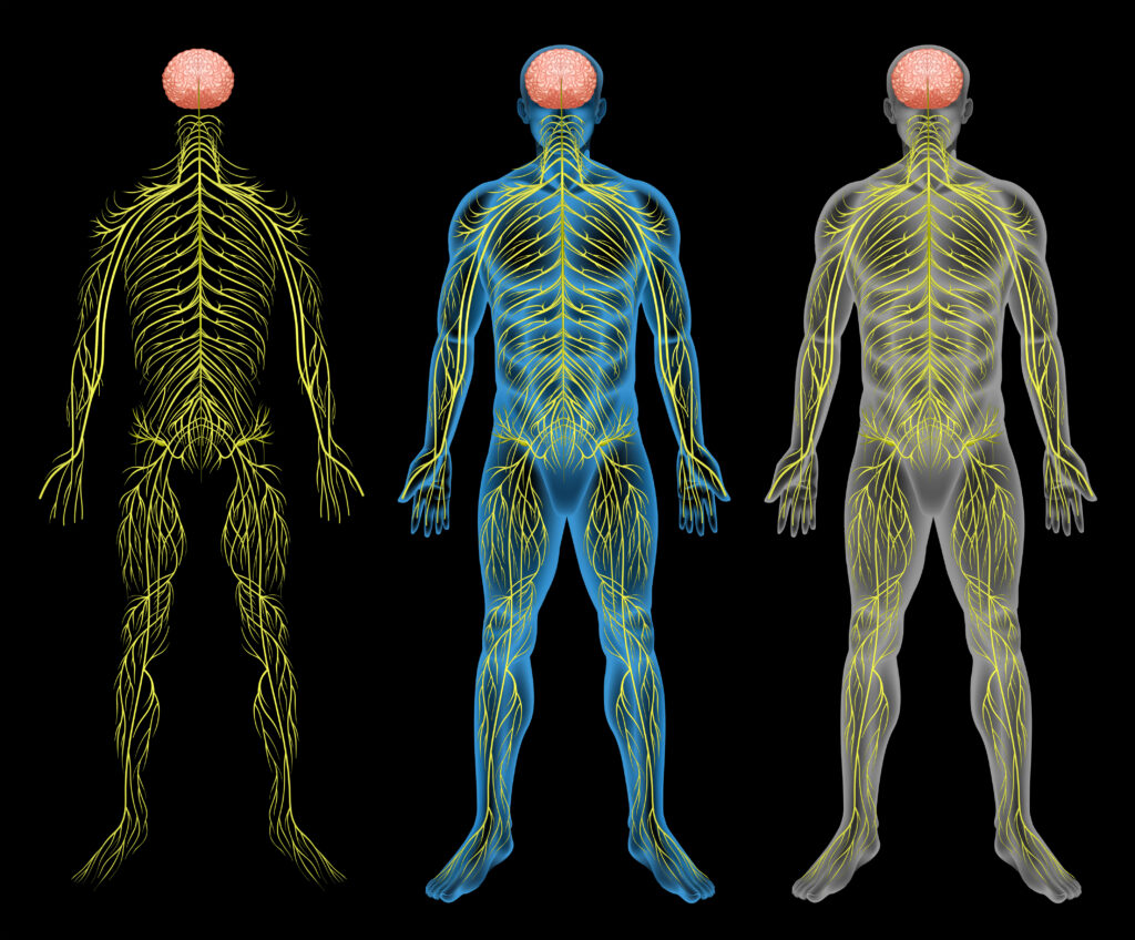
Peripheral nerves travel remarkable distances throughout the body, crossing multiple joints, passing through numerous anatomical tunnels, and interacting with muscles, fascia, and connective tissues along the way. Because of this complexity, nerve irritation often mimics orthopedic disorders.
Understanding peripheral nerve anatomy is one of the most valuable skills an orthopedic clinician can develop.
Built for Movement
Unlike tendons, which are designed primarily to transmit muscular force, peripheral nerves are designed to move.
During normal daily activities, nerves:
- Slide
- Glide
- Elongate
- Shorten
- Twist
- Adapt to changing joint positions
For example, during full elbow flexion the median nerve may glide nearly a centimeter relative to surrounding tissues. The sciatic nerve may glide almost an inch at 60˚ of a straight leg raise. Similar excursions occur in the upper and lower extremity during walking, pushing, pulling and lifting activities.
This mobility protects against mechanical stress while allowing normal movement. Problems arise when this mobility becomes restricted.
Double crush syndrome
One of the most important principles in peripheral nerve anatomy is that nerves function as continuous structures, traversing pathways through the body. Double crush syndrome occurs when there is compression along two different points of the nerve and synergistically increase symptoms.
Consider the patient who has:
- Cervical foraminal narrowing
- Mild carpal tunnel syndrome
- Diabetes
Each condition alone may produce minimal symptoms. Together, however, they may significantly impair nerve function.
Assessing peripheral nerves
Traditional neurological examination evaluates: strength, sensation and reflexes. While essential, these tests do not fully assess neural movement.
Neurodynamic testing evaluates how peripheral nerves tolerate movement throughout their anatomical course.
Examples include:
- Upper Limb Neurodynamic Tests (ULNT)
- Straight Leg Raise
- Slump Test
- Femoral Nerve Tension Test
A positive neurodynamic test does not necessarily indicate nerve damage. Instead, it may reflect increased neural mechanosensitivity or reduced neural mobility.
Clinical interpretation always requires correlation with the patient’s history and examination.
Why does this matter?
When evaluating musculoskeletal pain, ask yourself:
- Does the symptom distribution follow a peripheral nerve?
- Is there numbness or tingling?
- Are symptoms influenced by neck or spinal position?
- Is there weakness beyond pain inhibition?
- Does neural loading reproduce symptoms?
- Is there more than one possible compression site?
These questions often shift the clinician from a purely orthopedic diagnosis to a more comprehensive neurological assessment.
Peripheral nerves are not static electrical wires. They are dynamic, mobile tissues that must glide, elongate, and adapt to movement throughout the body. Appreciating their anatomy helps clinicians distinguish neural disorders from musculoskeletal pathology and develop more accurate diagnoses and effective treatment plans.
We’ll spend the next few weeks reviewing the path of major nerves so that you can begin to think of assessment, treatment and movement pattern solutions to common peripheral nerve problems.
Join me next week as we take a deeper dive into the path of the median nerve.
Because nobody has time to be in pain.
Until next time…

Kind Regards,
MoveWell Academy
[email protected]