Muscle Minute TUESDAY – Meet the Semimembranosus
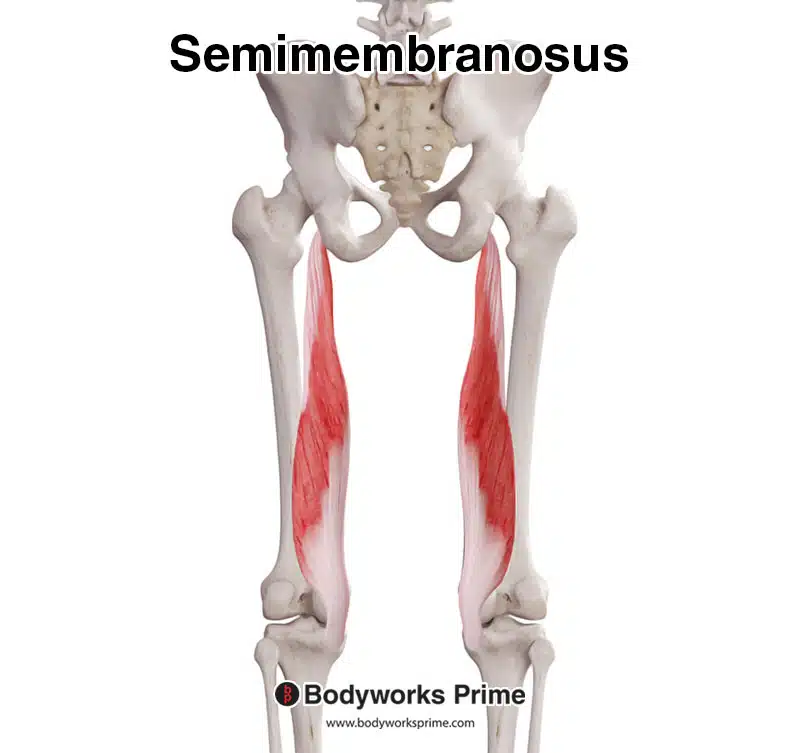
The hamstring muscle group is made up of three distinct muscles: semimbranosus, semitendinous and bicep femoris. The key word in that sentence is distinct. Though they tend to get lumped into one, clinically, they differ in structure and function. So I thought I’d start a mini-series on the hamstring muscles. Let’s take a closer look at semimembranosus (SM).
| Origin | lateral ischial tuberosity |
| Insertion | medial condyle of tibia |
| Action | hip extension, knee flexion, tibial internal rotation, dynamic meidal knee stabilizer (synergist of MCL) |
| Innervation | tibial n. (L5, S1, S2) |
| Antagonists | quadricep, bicep femoris |
The Real World Semimembranosus
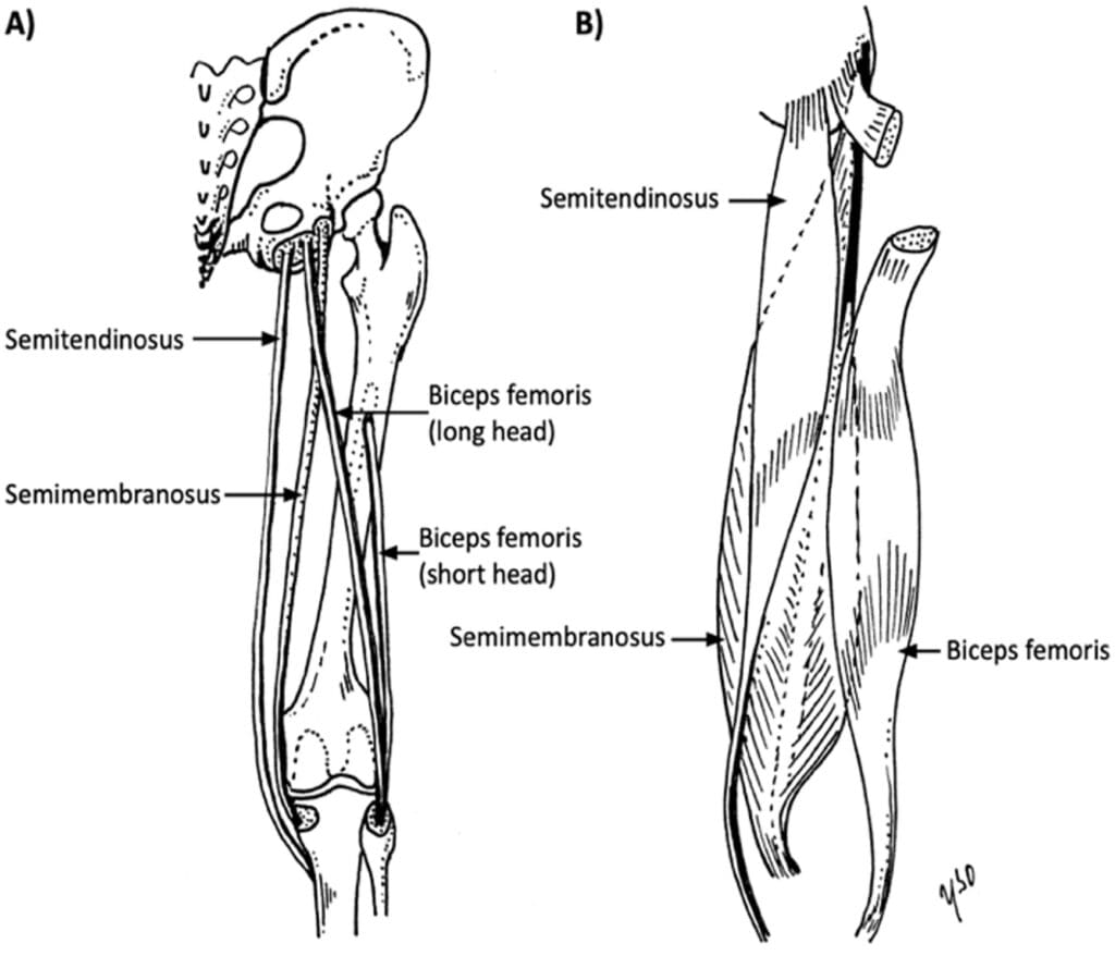
What makes this muscle unique? Of the two medial hamstrings, the distal tendon of SM lies deepest. Muscles oriented close to the joint are designed for stability and the SM is intimately located to the medial collateral ligament (MCL) of the knee, providing dynamic stabilization against valgus. The oblique popliteal ligament arises from the semimembranosus tendon providing posterior stability against knee valgus and extension.
The proximal tendon of the SM lies most lateral of the three hamstring tendons on the ischial tuberosity and the muscle belly is the largest of the three.
The diagonal orientation of this muscle lends itself to greater valgus control.
Here’s a short list of why this matters:
- Semimembranosus is the first line of defense against MCL tears of the knee
- Semimembranosus will be more susceptible to tearing with rapid hip flexion with the knee extended and the foot pronated
- Over pronators will be more likely to tear semimembranosus than the other hamstring muscles
- Semimembranosus may be placed in a long/weak position in someone who toes-out(tibial external rotation) when they walk or run
- Training or rehabilitation of the semimembranosis must include medial stabilizationactivities of the knee
- Training or rehabilitation of the semimembranosis should include strengthening of gluteus medius and restoration of normal ankle dorsiflexion, the two most common causes of over pronation.
Conventional thinking says: All the hamstrings extend the hip and flex the knee.
Real World Thinking says: Though the hamstrings have some function in common, each hamstring muscle is distinct in its function and location. The semimembranosus is a prime medial knee stabilizer, susceptible to injury in over pronation scenarios. It is the longest and largest of the hamstring muscle group and the second most prone to injury (after the bicep femoris). Rehabilitation of the semimembranosus should include stabilization against knee valgus.
Because nobody has time to be in pain.
Until next time…

Kind Regards,
MoveWell Academy
[email protected]