
Anatomy TUESDAY – The Coracoid Process
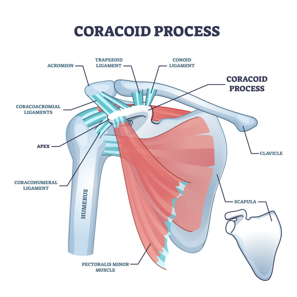
The coracoid process could be called the “busiest little piece of bone in the shoulder.” Before we discuss it, let’s see if you can find it.
Palpate the concave (lateral) portion of your clavicle with one hand. Drop your palpating fingers about an inch. Then extend and externally rotate your shoulder. You should feel the tip of the coracoid process, a hook-shaped projection extending from the scapula.
It isn’t particularly large, and it doesn’t form one of the primary joint surfaces of the shoulder. Yet an impressive number of muscles and ligaments converge on this small piece of bone.
The coracoid is essentially an anatomical anchor point and crossroads. Understanding what attaches to it helps explain how the scapula, clavicle and humerus work together—and why this little structure can be clinically important.
A Muscular Meeting Place
Three muscles attach directly to the coracoid process: the pectoralis minor, short head of the biceps brachii and coracobrachialis.
The pectoralis minor originates from the anterior surfaces of approximately ribs 3–5 and inserts onto the medial border and superior surface of the coracoid process. Because its other attachments are on the ribs, contraction of the pectoralis minor can pull the coracoid—and therefore the scapula—anteriorly and inferiorly around the thorax. It contributes to scapular protraction, downward rotation and anterior tilt.
This becomes particularly interesting clinically. A shortened or highly active pectoralis minor can be associated with altered resting scapular position and altered scapular movement. That doesn’t mean a “tight pec minor” automatically causes shoulder pain, but its anatomy gives us a good reason to consider it when evaluating scapular mechanics.
Two other muscles share a common attachment near the tip of the coracoid: the short head of the biceps and coracobrachialis. Their combined tendon is sometimes referred to as the conjoint tendon.
The short head of the biceps crosses both the shoulder and elbow, while the coracobrachialis runs from the coracoid to the medial humerus, flexing the shoulder. Together, these structures create an interesting muscular connection between the scapula and upper arm.
The Coracoid Is Also a Ligament Hub
Muscles are only half the story.
Several important ligaments attach to the coracoid, including the coracoacromial, coracoclavicular and coracohumeral ligaments.
The coracoacromial ligament runs between the coracoid and acromion, creating the coracoacromial arch over the humeral head. This arch forms part of the superior boundary of the subacromial region and is often an area of impingement in cases of faulty scapular positioning or mechanics.
The coracoclavicular ligament connects the coracoid to the clavicle and consists of two components: the conoid and trapezoid ligaments. The conoid checks tipping of the scapula while the trapezoid checks winging.
This relationship is especially important because the scapula doesn’t move independently of the clavicle. During arm elevation, movement occurs at the sternoclavicular and acromioclavicular joints as the scapula upwardly rotates, posteriorly tilts and externally rotates. The coracoclavicular ligaments help guide and constrain this coordinated motion.
The coracohumeral ligament extends from the coracoid toward the greater and lesser tuberosities of the humerus and blends with structures around the rotator interval. It contributes to glenohumeral stability and restraint of humeral translation depending on shoulder position.
So this small projection of bone participates indirectly in both scapular mechanics and glenohumeral stability.
The Coracoid, Pec Minor and Brachial Plexus
The anatomy becomes even more interesting when we look underneath the pectoralis minor.
The cords of the brachial plexus are actually named according to their relationship to the second part of the axillary artery—and this region lies deep and medial to the coracoid/pectoralis-minor complex. The coracoid therefore provides clinicians with a useful surface landmark for understanding deeper neurovascular anatomy.
The musculocutaneous nerve is another great anatomical pearl. After arising from the lateral cord of the brachial plexus, it typically pierces the coracobrachialis before traveling between the biceps and brachialis.
Think about that arrangement: a muscle originating on the coracoid is actually pierced by a major peripheral nerve.
This is exactly the kind of anatomical relationship that turns memorized anatomy into clinically useful anatomy.
Why Should a Clinician Care?
The coracoid is an excellent palpation landmark during shoulder examination. Once you locate it, you have a reference point for the pectoralis minor, conjoint tendon, clavicle, acromioclavicular region and the brachial plexus.
Tenderness around the coracoid can have multiple potential sources. Symptoms may involve the muscular attachments, adjacent soft tissues, or structures in the surrounding anterior shoulder. Location alone therefore doesn’t provide a diagnosis—but knowing the anatomy helps develop a much better differential.
The coracoid also reminds us why treating the shoulder as simply a ball-and-socket joint is inadequate.
Movement of the arm depends on coordinated motion between the humerus, scapula, clavicle and thorax. The coracoid sits almost directly in the middle of that system, providing attachments that connect the scapula to the ribs, clavicle and humerus.
For such a small piece of bone, it has an enormous amount of responsibility.
The coracoid doesn’t create much movement itself. Instead, it serves as a strategic anchor where muscles and ligaments connect the scapula to the ribs, clavicle and humerus—making it one of the most functionally important landmarks in the shoulder.
Consider the coracoid.
Because nobody has time to be in pain.
Until next time…
Kind Regards,
MoveWell Academy
[email protected]