Anatomy TUESDAY – Palpation Series (Part 7) – Medial Ankle
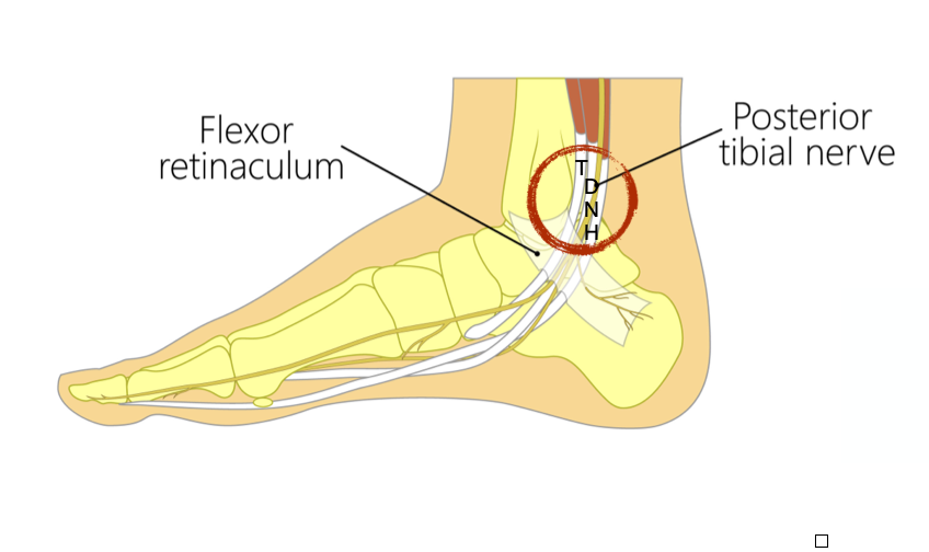
If you’ve ever been in an anatomy class, you are familiar with the mnemonic: Tom, Dick ANd Harry, used to remember the tendons and structures on the medial side of the ankle.
- Tom = Tibialis Posterior
- Dick = Flexor Digitorum Longus
- AN = Tibial artery and nerve
- Harry = Flexor Hallucis Longus
Check out our medial ankle palpation video and follow along. If you place your index finger on the medial malleolus and then fall off of it posteriorly, the width of your finger lands on the area circled above and encompasses all of the above structures. If you slide your finger to just inferior to the tip of the medial malleolus, you are now in the area of the flexor retinaculum, the ligament under which all of those structures pass.
Why does this matter?
Compression of tibial nerve in this region may cause tarsal tunnel syndrome and result in pain along the medial side of the foot and ankle region.
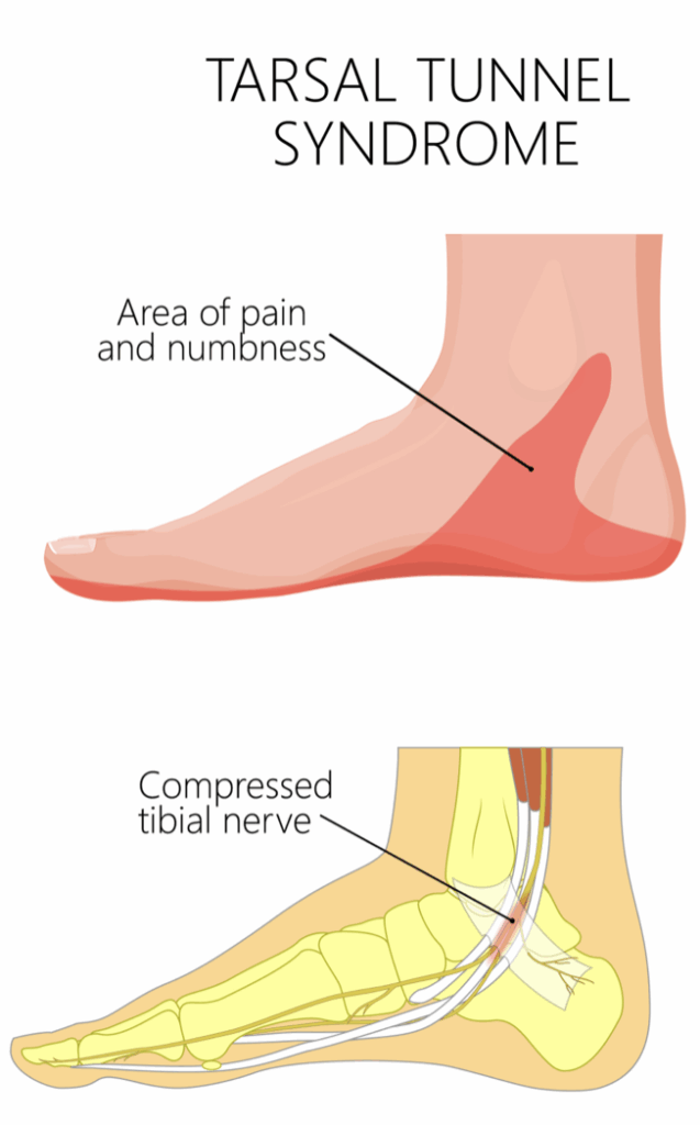
But what causes that compression? Is it a tightening of the retinaculum? No. It is chronic swelling of the tendons surrounding the nerve. And what causes the chronic swelling of Tom, Dick ANd Harry? Over pronation.
And what are the two most common causes of over pronation? (hint: it isn’t the lack of an orthotic)
- Weak gluteus medius
- Lack of ankle dorsiflexion (from tight gastrocnemius or an ankle jam)
Pain with palpation of the medial ankle structures could tell us all of this?! Yes.
So, take your thumb or index finger and palpate the inch behind your medial malleolus. Is it tender? Is it more tender on one side than the other? If so, you may have just discovered you over pronate on that side. Here’s some exercises to help you out if this is the case. (click on the link to download a copy)

Because nobody has time to be in pain.
Until next time…

Kind Regards,
MoveWell Academy
[email protected]