

Anatomy TUESDAY – The Sciatic Nerve and the Straight Leg Raise (SLR)
A patient lies on their back while the clinician performs a straight leg raise. At about 60 degrees, the patient reports tightness behind the thigh.
“That’s my hamstring. It’s always tight.”
Then the clinician adds ankle dorsiflexion.
Suddenly, the sensation becomes stronger or travels farther down the leg.
The hamstring didn’t suddenly become shorter.
The mechanical load on the nervous system changed.
The sciatic nerve isn’t simply a cable carrying signals from the spine to the leg. It is a living, mobile tissue that must glide and adapt as the hip, knee, ankle, and spine move.
Follow the Sciatic Nerve
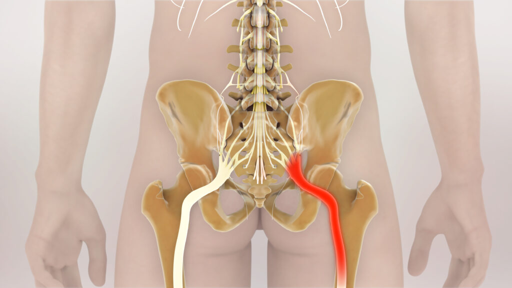
The sciatic nerve is the largest peripheral nerve in the human body. It arises from the L4–S3 nerve roots through the lumbosacral plexus.
It exits the pelvis through the greater sciatic foramen, typically passing beneath the piriformis, and travels through the deep gluteal region before descending along the posterior thigh.
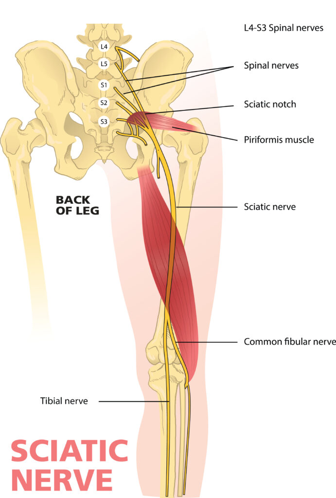
The nerve actually contains two major neural components traveling together:
- Tibial division
- Common fibular (peroneal) division
Near the popliteal region—although the level of division varies—the two separate into the tibial and common fibular nerves.
From the lumbar spine to the foot, this neural pathway crosses multiple joints.
That creates an interesting mechanical problem.
How does a nerve travel from the spine to the foot when the distance between those two points constantly changes?
The answer is movement.
Peripheral nerves are designed to accommodate movement through a combination of excursion, elongation and changes in their relationship to surrounding tissues.
Consider what happens when you walk.
As one leg swings forward:
- The hip flexes
- The knee extends
- The ankle dorsiflexes
- The pelvis rotates
- The lumbar spine rotates
If the sciatic nerve were rigidly attached to every structure it passed, normal movement would place enormous mechanical stress on it. Instead, the nerve moves relative to its surrounding tissues. This ability to glide is part of normal neurodynamics.
Is That Really a Tight Hamstring?

A patient may report posterior thigh tightness during:
- Toe touching
- Straight leg raising
- Long sitting
- Running
- Hamstring stretching
It is easy to assume the hamstring is simply short. But the sensation of “tightness” doesn’t identify the tissue responsible. Muscle, fascia and neural tissues are all being mechanically affected during these movements.
One simple modification can provide an important clue.
Dorsiflex the ankle.
The hamstrings don’t cross the ankle. Therefore, ankle dorsiflexion produces essentially no meaningful additional lengthening of the hamstring muscle-tendon unit.
But it does alter mechanical loading of the neural system, particularly through the tibial nerve and its continuity with the sciatic nerve.
If ankle dorsiflexion significantly changes the patient’s posterior thigh or leg symptoms, the clinician has changed neural loading without meaningfully changing hamstring length.
That is called structural differentiation.
It provides evidence that neural tissues may be contributing to the patient’s response.
Stretching Isn’t Always the Answer
If a patient repeatedly describes their hamstrings as tight, the natural response is often to stretch them harder. But if neural mechanosensitivity is contributing to that sensation, aggressive stretching may simply increase neural loading.
This is where nerve sliders can be useful.
A slider increases mechanical loading at one end of the neural pathway while simultaneously reducing it at the other.
For example, during a seated sciatic nerve slider, the patient might:
Extend the knee while extending the neck, then flex the knee while flexing the neck.
The goal is to encourage neural excursion without creating large amounts of overall neural strain.
Here’s a great video on sciatic nerve flossing. (And the guy has a great accent that makes you want to listen).
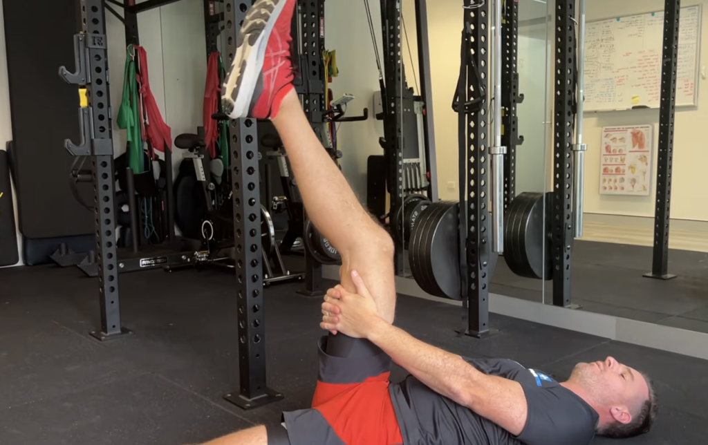
A tensioner, in contrast, progressively loads both ends of the neural pathway simultaneously. (For the sciatic nerve, this is hip flexion, knee extension, ankle dorsiflexion)
Tensioners create greater mechanical demand and are generally more appropriate once irritability has decreased and greater loading is indicated.
A useful clinical distinction is:
Sliders encourage neural excursion. Tensioners progressively increase neural loading.
Why does this matter?
The sciatic nerve is not a stationary cable running down the leg. It is a mobile tissue that must accommodate movement from the lumbar spine all the way to the foot.
Hip flexion and knee extension progressively increase mechanical demand on this neural pathway, which is why the straight leg raise provides information about much more than hamstring flexibility.
Adding ankle dorsiflexion makes the test even more informative because it increases neural loading without meaningfully increasing hamstring length.
The next time a patient says, “My hamstrings are always tight,” don’t immediately assume the muscle needs more stretching.
Ask:
Does the sensation change when I alter neural load?
Sometimes what feels like a tight muscle isn’t primarily a muscle flexibility problem at all.
It may be the nervous system telling you that it doesn’t currently tolerate that combination of movement and load. Try sciatic nerve flossing.
Because nobody has time to be in pain.
Until next time…

Kind Regards,
MoveWell Academy
[email protected]