Muscle Minute TUESDAY – Meet the Medial Collateral Ligament (Knee)
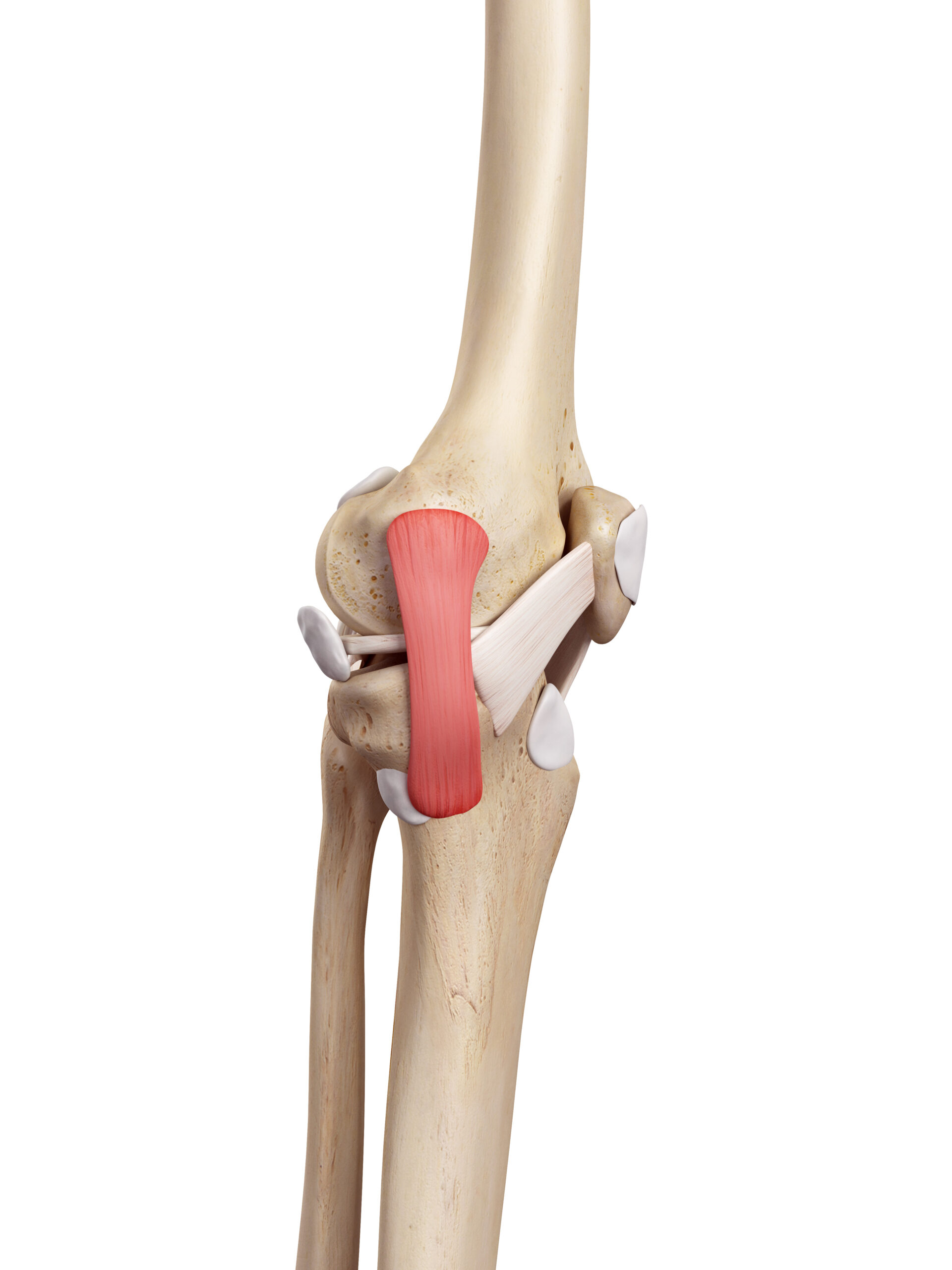
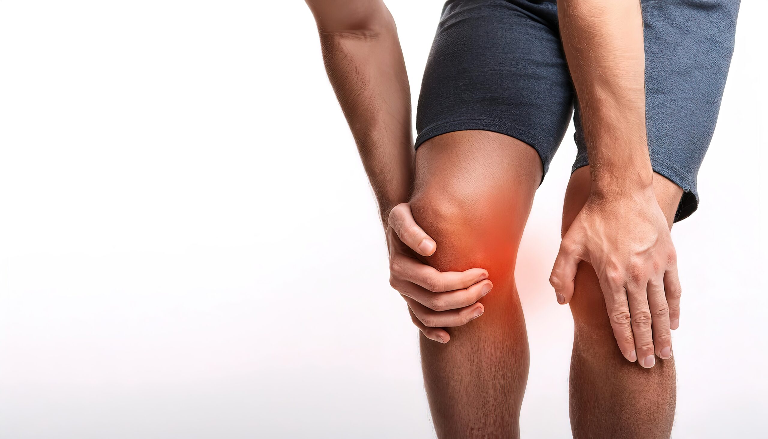
The medial collateral ligament (MCL) of the knee is the most commonly injured knee ligament involved 7.9% of all athletic knee injuries according to a 10-year observational study. It has two divisions: superficial and deep, attaching 1.2 cm and 6 cm below the medial tibial plateau respectively. The deep fibers are thought to resist excessive valgus while the superficial distal fibers are thought to prevent excessive internal/external rotation of the tibia.
Recently, I have had two cases where the MCL was injured in an overuse scenario: one after running 6 miles and another after prolonged standing and with stair descending.
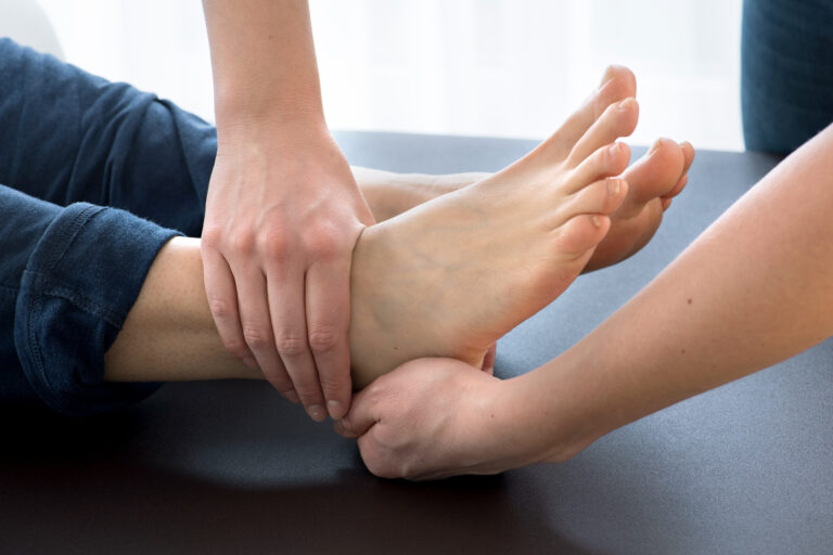
Both cases exhibited typical symptoms of a ligament tear: pain with palpation of the MCL and pain with valgus stress testing.
Both cases demonstrated a negative valgus stress test following release of one or more of the following muscles: medial gastrocnemius, adductor magnus and psoas major.
Let’s break those muscles down:
| Medial gastrocnemius | plantarflexion and supination of the ankle |
| Adductor magnus | adduction and external rotation of the femur |
| Psoas major | flexion and external rotation of the femur |
Real World MCL Relationships
What do any of these muscles have to do with knee valgus? I asked myself the same question after treatment yielded a negative valgus test and here’s what I came up with:
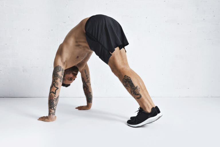
- Medial gastrocnemius – tightness would limit dorsiflexion, causing a toe-out compensation (ER of the tibia) during squatting and stair ambulation activities, increasing knee valgus
- Adductor magnus – tightness would cause adduction of the femur, increasing valgus of the knee and/or external rotation causing relative internal rotation of the tibia.
- Psoas major – tightness would cause hip external rotation, creating a relative internal rotation of the tibia and/or contributing to toe-out posture.
This discovery gives us major insight into specific exercises for cases like this:
- Strengthen gluteus medius to decrease excessive femoral adduction and counteract adductor magnus TrP.
- Increase ankle dorsiflexion to prevent toe-out compensation. (downward dog, gravity drop, BOSU rockers, long stride walking)
- Increase hip internal rotation. (lateral low row, pigeon pose, carioca stepping)
Conventional thinking says: The MCL is torn with rapid valgus stresses of the knee.
Real World Thinking asks: The MCL may also be placed on excessive tension with over pronation scenarios causing a toe-out posture, even without traumatic injury. In these cases, valgus instability may be altered immediately by releasing associated trigger points. It’s worth investigation.
Because nobody has time to be in pain.
Until next time…

Kind Regards,
MoveWell Academy
[email protected]